lmnature · Molecular biology · Page mb14
DHFR coordinate geometry with MTX and NDP
Media on this page: text, diagram, video and live animation are available.
The pinned RCSB 3DFR PDB file reports X-RAY DIFFRACTION at 1.70 Å resolution. Its DBREF row maps chain A residues 1–162 to UniProt P00381. The coordinate rows number MTX as chain A residue 164 and NDP as chain A residue 163. The deposited asymmetric unit contains chain A with these HETATM groups. The PDB header marks the protein as engineered and describes the author-designated biological assembly as monomeric for chain A. COMPND identifies DIHYDROFOLATE REDUCTASE from Lactobacillus casei. HETNAM names MTX METHOTREXATE and NDP NADPH DIHYDRO-NICOTINAMIDE-ADENINE-DINUCLEOTIDE PHOSPHATE. RCSB PDB 3DFR (CC0); primary paper citation: Bolin et al., J. Biol. Chem. volume 257 (1982), PMID 6815178.
What was measured
Each value below is the Euclidean separation between two heavy-atom coordinate positions in the pinned PDB. The second decimal is arithmetic rounding, not an experimental precision statement.
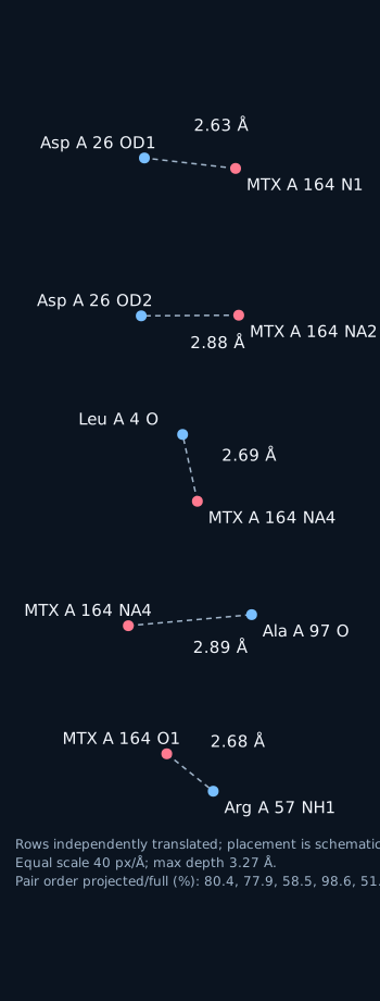
- MTX A 164 N1 to Asp A 26 OD1: 2.63 Å. B-factors: 12.40 and 10.10 Ų.
- MTX A 164 NA2 to Asp A 26 OD2: 2.88 Å. B-factors: 9.00 and 13.30 Ų.
- MTX A 164 NA4 to Leu A 4 O: 2.69 Å. B-factors: 9.10 and 11.00 Ų.
- MTX A 164 NA4 to Ala A 97 O: 2.89 Å. B-factors: 9.10 and 14.10 Ų.
- MTX A 164 O1 to Arg A 57 NH1: 2.68 Å. B-factors: 22.80 and 14.30 Ų.
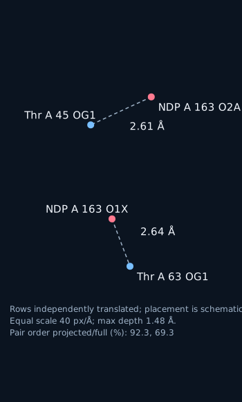
- NDP A 163 O2A to Thr A 45 OG1: 2.61 Å. B-factors: 13.90 and 18.30 Ų.
- NDP A 163 O1X to Thr A 63 OG1: 2.64 Å. B-factors: 18.30 and 19.60 Ų.
The 3.0 Å native scene contains 19 distance-contact lines, including all 7 listed pairs. Each selected pair appears as a scene contact entity in every frame. The numbers are coordinate measurements; no enzyme-process interpretation is assigned.
MTX coordinate pairs
NDP coordinate pairs
PDB records and coordinate limits
The fixed-column file has 1,294 ATOM rows and 345 HETATM rows, including 33 MTX atoms, 48 NDP atoms, and 264 water atoms; 1,375 non-water heavy atoms are retained. It has 1 coordinate model, 81 CONECT rows, 36 SITE rows, and 0 LINK rows. CONECT rows list atom connections within MTX and NDP; none joins a protein atom to either group. These are deposited file annotations, separate from the coordinate measurements. AltLoc alternatives: 0; hydrogen/deuterium coordinate rows: 0; ligand occupancy values: MTX 1.00; NDP 1.00. REMARK 470 lists LYS A 51: CG, CD, CE, NZ as absent. RCSB PDB 3DFR.
What these coordinates do and do not show
This is one static coordinate model. It does not show atoms moving over time or a reaction sequence. The coordinate file contains no hydrogen positions; the heavy-atom separations alone do not locate hydrogen positions. No enzyme function or reaction effect is assigned to any measured distance. RCSB PDB 3DFR; PMID 6815178.
Video: fixed coordinates, rotating camera
Live: orbit it yourself
Heavy-atom distances are geometric measurements, not inferred bonds. A single static snapshot; no atom moves.
Papers the corpus links to this protein
The corpus links 8 papers across 1 UniProt entry (P00381); per-protein counts: P00381: 8. These papers are cross-referenced by the corpus and are not necessarily about this structure.
Titles, journal and year load from Europe PMC when JavaScript is available.