lmnature · Molecular biology · Page 12
Trypsin and BPTI: measured distances across the 2PTC interface
Media on this page: text, diagram, video and live animation are available.
The pinned RCSB 2PTC file records a 1.90 Å X-ray structure with trypsin on chain E and BPTI on chain I. RCSB PDB 2PTC identifies the coordinate entry and its metadata. The PDB-cited paper by Marquart et al. is linked by its verified primary-source DOI, doi:10.1107/S010876818300275X; the DOI was verified against the primary IUCr article record.
What was measured
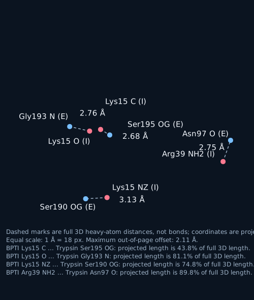
Each value is the Euclidean distance between the named heavy-atom coordinates in the pinned file. These four E-I pairs were selected because each has a corresponding contact entity in the native video scene at 3.5 Å. Distances are reported to two decimals; the second decimal is arithmetic rounding, not coordinate precision.
- BPTI Lys15 C ... Trypsin Ser195 OG: 2.68 Å. B-factors: 16.55 and 9.54 Ų.
- BPTI Lys15 O ... Trypsin Gly193 N: 2.76 Å. B-factors: 16.55 and 18.28 Ų.
- BPTI Lys15 NZ ... Trypsin Ser190 OG: 3.13 Å. B-factors: 14.78 and 15.42 Ų.
- BPTI Arg39 NH2 ... Trypsin Asn97 O: 2.75 Å. B-factors: 35.06 and 24.81 Ų.
What the file records
The PDB has 2083 ATOM records in chains E and I and 158 HETATM records: 157 waters plus one calcium atom, CA E 462 (serial 2086, occupancy 0.50, B-factor 14.17 Ų). The selected interface pairs have no matching LINK or CONECT records. The file has 6 LINK, 26 CONECT, and 2 SITE records, including SITE annotation AC1; none of the selected E-I pairs appears in those records. Selected-atom occupancies are 1.00. The two protein chains are marked ENGINEERED in COMPND. The file has no alternate locations and no hydrogen atoms. Atom labels use the deposited PDB chain and residue numbering. These details are from the pinned RCSB PDB 2PTC.
The asymmetric-unit coordinates contain chains E and I. PDB REMARK 350 labels BIOMOLECULE 1 an author-determined dimer; it also lists a separate software-generated octamer assembly. This page uses the raw asymmetric-unit coordinates and adds no symmetry copies. Source: RCSB PDB 2PTC.
What the video shows and does not show
At a 3.5 Å cutoff, all four selected E-I distances listed above have matching native-scene contact entities. The scene also includes calcium-to-chain-E lines; these are separate from the selected interface measurements. At a 3.0 Å cutoff, the 3.13 Å Lys15 NZ ... Ser190 OG pair is above the cutoff and is not drawn. A static coordinate model does not show molecular motion or establish a functional outcome. This page reports distances only.
Video: fixed 2PTC coordinates, rotating view
Live: orbit it yourself
Heavy-atom distances are geometric measurements, not inferred bonds. A single static snapshot; no atom moves.
Papers the corpus links to this protein
The corpus links 22 papers across 2 UniProt entries (P00760, P00974); per-protein counts: P00760: 11; P00974: 11. These papers are cross-referenced by the corpus and are not necessarily about this structure.
Titles, journal and year load from Europe PMC when JavaScript is available.