lmnature · Molecular biology · Page mb04
Lysozyme cleft and (NAG)3 geometry
Media on this page: text, diagram, video and live animation are available.
The pinned RCSB 1HEW coordinates are an X-ray structure at 1.75 Å resolution. Chain A is hen egg-white lysozyme; chain B contains 3 NAG residues. The PDB author-designated biological unit is monomeric, with chains A and B listed under an identity transform. The file marks the protein as engineered. Its 1,001 ATOM records cover 129 residues; 146 HETATM records include 43 NAG atoms and 103 water atoms. The renderer retains the protein and NAG (1,044 atoms) and omits water. RCSB PDB 1HEW, CC0; Cheetham, Artymiuk & Phillips, Journal of Molecular Biology 224, 613–628 (1992), DOI 10.1016/0022-2836(92)90548-X; PMID 1569548.
What was measured
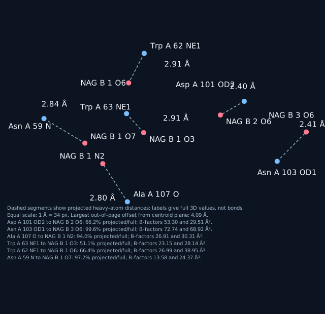
Each value is a Euclidean distance between two heavy-atom coordinates in the pinned file. The second decimal is arithmetic rounding, not experimental precision.
- Asp A 101 OD2 to NAG B 2 O6: 2.40 Å. B-factors: 53.30 and 29.51 Ų.
- Asn A 103 OD1 to NAG B 3 O6: 2.41 Å. B-factors: 72.74 and 68.92 Ų.
- Ala A 107 O to NAG B 1 N2: 2.80 Å. B-factors: 26.91 and 30.31 Ų.
- Trp A 63 NE1 to NAG B 1 O3: 2.91 Å. B-factors: 23.15 and 28.14 Ų.
- Trp A 62 NE1 to NAG B 1 O6: 2.91 Å. B-factors: 26.99 and 38.95 Ų.
- Asn A 59 N to NAG B 1 O7: 2.84 Å. B-factors: 13.58 and 24.37 Ų.
At a 4.0 Å renderer cutoff, 51 protein-to-NAG distance lines are included; at 3.0 Å, 7 remain. The 3.0 Å set includes Asp A 101 OD2 to NAG B 2 C6 at 2.90 Å, an oxygen-to-carbon pair, so the cutoff does not classify all 7 as polar. The deposited paper describes tri-N-acetylchitotriose as an inhibitor at partial occupancy, while all 43 NAG coordinate rows list occupancy 1.00.
PDB records
The file has 2 LINK rows for NAG B 1 O4 to NAG B 2 C1 (recorded value 1.48 Å) and NAG B 2 O4 to NAG B 3 C1 (1.45 Å); these are file records, not measurements made for the list above. Its 51 CONECT rows encode 4 protein disulfide pairs and NAG connectivity; none connect protein to NAG. The pinned file has no SITE records. All alternate-location fields are blank, and the file contains no hydrogen or deuterium atoms. REMARK 350 identifies the biological unit as monomeric; the deposited asymmetric-unit coordinates already include the listed chains. These record statements refer to RCSB PDB 1HEW, not an inference from the plotted distances.
What this view does and does not show
The diagram and video focus on the six selected protein-to-NAG distances. They do not show the Glu A 35 or Asp A 52 sidechains: the closest coordinates are 5.95 Å and 4.03 Å from NAG, respectively, outside the renderer’s 4.0 Å cutoff. Those residues are not labeled or drawn as sidechains in either view. Cheetham et al. (1992), PMID 1569548.
Limits: one static coordinate model; it shows no movement or reaction. The file does not include hydrogens, so these heavy-atom separations alone do not establish a hydrogen-mediated interaction. The paper and file occupancy fields differ as described above.
Video: fixed coordinates, rotating camera
Live: orbit it yourself
Heavy-atom distances are geometric measurements, not inferred bonds. A single static snapshot; no atom moves.
Papers the corpus links to this protein
The corpus links 17 papers across 1 UniProt entry (P00698); per-protein counts: P00698: 17. These papers are cross-referenced by the corpus and are not necessarily about this structure.
Titles, journal and year load from Europe PMC when JavaScript is available.