lmnature · Molecular biology · Page 1
Myoglobin holds one oxygen next to one iron
Muscle needs oxygen faster than blood can hand it over. Myoglobin is a single chain of 153 amino acids that sits inside muscle cells and keeps oxygen on hand. It does this with help: tucked into a pocket in the folded chain is a flat, iron-containing ring called heme, and an oxygen molecule (O2) is held beside that iron.
The structure on this page is 1MBO, sperm-whale oxy-myoglobin solved by X-ray crystallography (Phillips 1980, J. Mol. Biol. 142, 531–554; RCSB PDB 1MBO, CC0). The same protein is described in the lmnature corpus as UniProt P02185.
What was measured
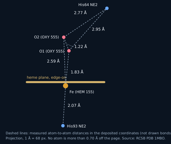
Everything in this section is a distance between two atom positions in that deposited file, computed by this page’s build script. The file records connections (LINK and CONECT lines) from the heme iron to the nitrogen NE2 of histidine 93 (the proximal histidine), to the four nitrogens of the heme ring, and to both oxygen atoms of the O2 molecule (residue OXY 555). Measured: His93 NE2 is 2.07 Å from the iron; the ring nitrogens are 1.90–2.00 Å from it; the nearer oxygen atom is 1.83 Å from the iron and the farther one 2.59 Å; the two oxygens are 1.22 Å apart. On the other face of the heme, the NE2 nitrogen of histidine 64 (the distal histidine) is 2.95 Å from the nearer oxygen and 2.77 Å from the farther one.
What this does and does not tell you
Interpretation, kept separate from the measurement: a 1.83 Å iron–oxygen distance is what you would expect if the oxygen is bound to the iron, and the distal histidine is positioned where it could hold the oxygen by a hydrogen bond. A distance alone does not prove either. A hydrogen bond also depends on the hydrogen position and the angle, which this structure does not show, and the file records no hydrogen atoms. The links listed in the file are the depositors’ declared connections, not something this page infers.
Precision: the data are at 1.60 Šresolution and the two oxygen atoms have B-factors of 20.66 and 19.61 Ų, so they are less sharply located than the protein core. Read the second decimal place as arithmetic, not as the precision of the measurement.
Not shown: the structure is a single static snapshot. It does not show oxygen arriving or leaving, and nothing on this page is a simulation.
Video: one turn around the whole protein
This MP4 is rendered by lmvideo from the same LMCode source that drives the live view below.
Live: orbit it yourself
Drag to orbit, scroll to zoom, click an atom to see its identifier. The frames are computed in your browser. Green lines are heavy-atom distances at or under a cutoff, not bonds; the solid sticks between hetero atoms (for example iron–oxygen) come from the file’s own CONECT records. Exact views: 4 Å (all contacts within 4 Å) · 3 Å: the contacts drawn then include the two histidine 64 distances (2.77 and 2.95 Å), and also others.
Papers the corpus links to this protein
The corpus links 8 papers across 1 UniProt entry (P02185); per-protein counts: P02185: 8. These papers are cross-referenced by the corpus and are not necessarily about this structure.
Titles, journal and year load from Europe PMC when JavaScript is available.